Harnessing the power of AI in research

Article by: Lisa Lopez-Snyder
Originally Published
How powerful can artificial intelligence (AI) be as an infectious disease research tool for pathologists?
A team of experts from The Ohio State University College of Veterinary Medicine’s Combined Pathology Training/ Graduate Program are at the cutting edge of answering that question. Led by Michael Oglesbee, DVM, PhD, DACVP, professor of veterinary biosciences and director of Ohio State’s Infectious Diseases Institute, the program is breaking new ground by leveraging AI to revolutionize pathology
training and diagnostics in infectious disease research.

At the forefront of this initiative is Kara Corps, DVM, PhD, DACVP, assistant clinical professor and director of the Comparative Pathology & Digital Imaging Shared Resource, who serves as the project’s principal investigator.
“Developing AI-based models that can perform this complex work requires teaching the AI to accurately identify and integrate complex data,” Corps explains. “It’s a multistep process, and much effort goes into building the pipeline before we can deploy these models for practical use.”
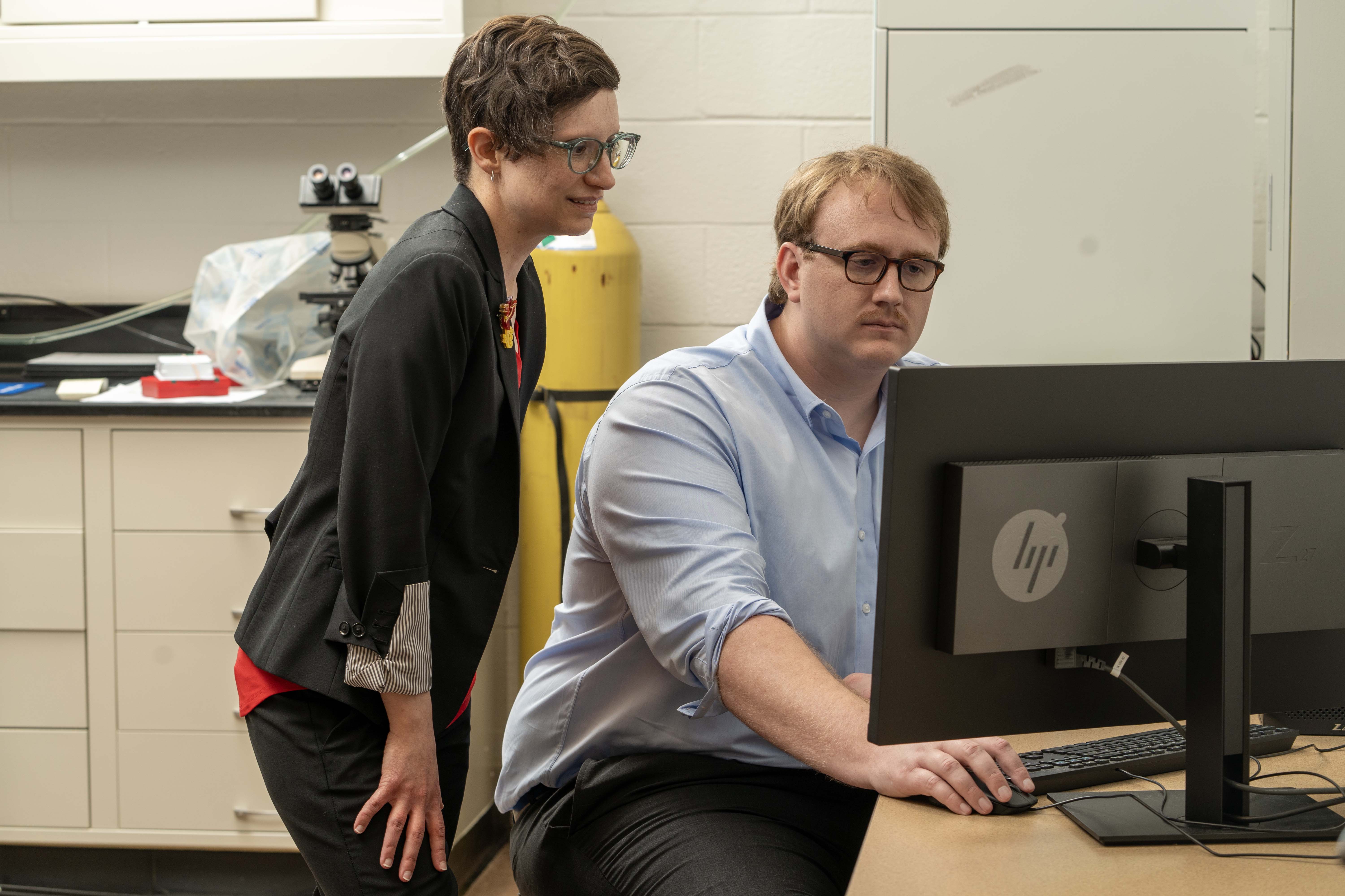
That’s where Sam Neal, DVM, a third-year clinical pathology trainee, comes in. Neal is doing the hands-on work, building and refining the AI models to advance infectious disease research.
Neal, an expert in AI-driven technology research, says the goal is to allow AI to augment the tedious tasks involved in research and “to allow us as pathologists to use our knowledge to elucidate the underlying mechanisms of disease.”
He says the project’s desired outcome is ultimately to provide knowledge on improving the prevention, diagnosis and treatment of infectious disease in animals and humans. Neal’s efforts exemplify the innovative spirit driving this project.
This ambitious initiative was made possible through a collaboration between Ohio State College of Veterinary Medicine and Charles River Laboratories (Charles River), whose partnership helped establish the fund supporting the program. The inital funding from Charles River marks the first step toward creating a sustainable, far-reaching program that equips future pathologists with the tools needed to lead advancements in AI-driven disease research.
The study’s three phases
The AI project has three phases. In the first phase, Neal is collecting glass slides of mice lung samples with infections from one of three key pathogens: SARSCoV2, the infectious agent of COVID 19; Bordetella bronchiseptica, a pathogen that causes widespread respiratory disease in dogs, pigs and cattle and emerging in humans; and Bordetella pertussis, the causative agent of whooping cough and a top 10 cause of infant mortality in the world.
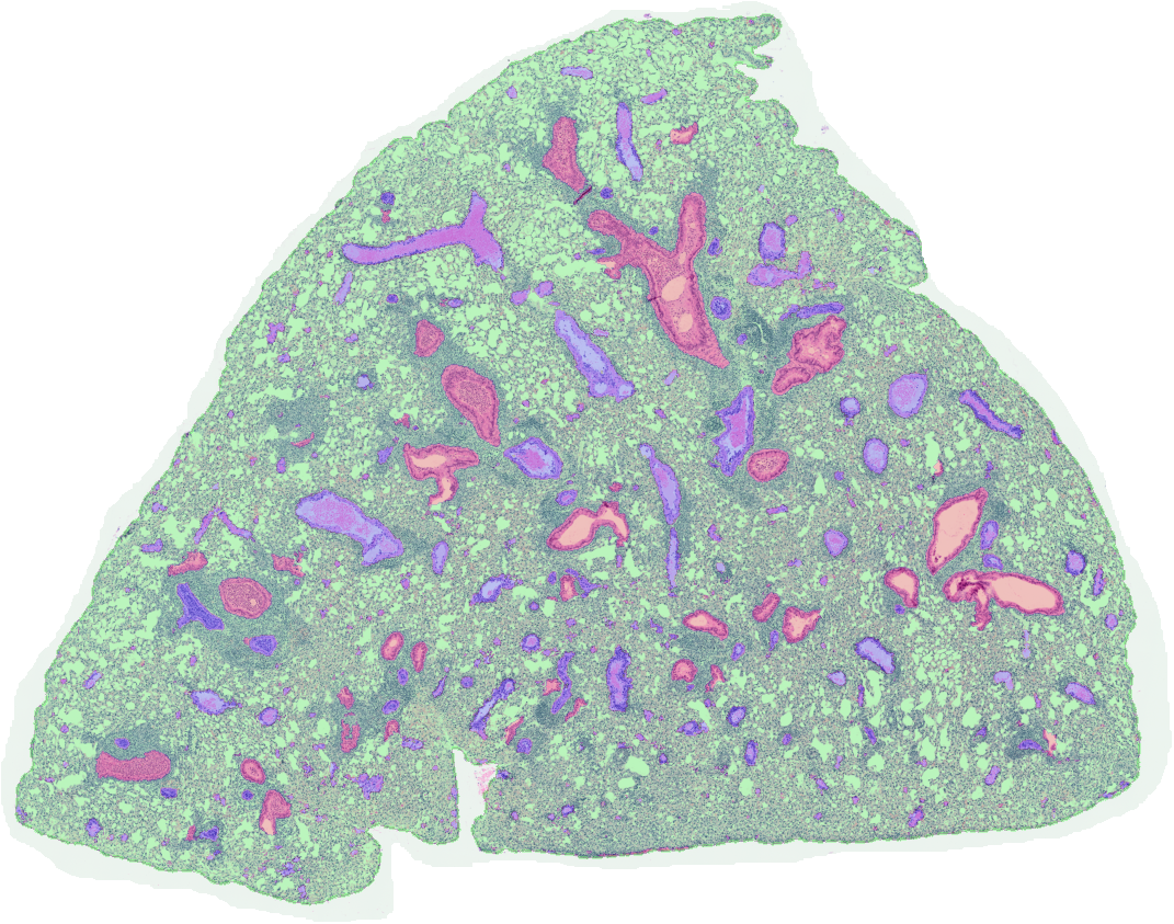
In the next phase, Neal is training the AI tool to identify and understand all aspects of pathology, including, for example, learning what an airway is, what a blood cell is, what cell structures are and how they’re spatially related to one another.
“The training phase is comprehensive,” says Corps. “It goes all the way from identifying the tissue to being able to identify individual cells in a variety of circumstances,” she says. “To do that properly, Dr. Neal must go through hundreds of slides to teach the AI how to identify and relate things appropriately, then add and train the AI with correlated data.”
The third phase involves testing and refining the tool. The ambitious goal is to develop a model for implementation by the collaborative pathology and research team by the end of 2024.
Making an impact
Oglesbee says Charles River’s support is significant for the future of biomedical research.
“Now we can really push the envelope and not only focus on the image itself but also explore how we can enhance our ability to interpret what’s going on when we fold in additional data streams addressing lung function, immune and infection status,” he says.

“For example, this tool may tell us something about the severity of a disease and what might be driving it. This is relevant—and important to know—as you develop treatments or vaccines. This is pushing the boundaries of what we have been trained to do traditionally,” says Oglesbee.
Dan Rudmann, DVM, PhD, DACVP, FIATP, was the senior director of Digital Toxicologic Pathology at Charles River at the time when Neal completed his veterinary internship there.
“Dr. Neal had a lot of promise as a leader and we had a shared interest in applying AI in a translational way, so it seemed like a big win for us to support his development as a young scientist,” he says.
Rudmann says Neal’s interest in new technology that uses AI to solve medical problems and help data have a more significant impact on human and veterinary patients led Charles River Laboratories to support his work.
Later, once Neal began his residency program, the company connected with Ohio State and “carved out a way to be involved in developing him as a leader,” says Rudmann, an investigative and toxicologic pathologist and translational medicine scientist who is now executive director of Toxicologic Pathology at Moderna.
Developing expertise in AI pathology
Michael Staup, PhD, associate scientific director of Digital Pathology at Charles River, says supporting Neal is exciting as the company seeks to recruit young pathologists like him to do this specialized work.
“The work we do in safety assessments is vital and relies upon trained and boarded veterinary pathologists, so being able to curate the best and the brightest and develop a relationship with them is the most important thing for us.”
He says the project is ambitious. “Right now, we are just trying to get the classifications down. Most AI-assisted image analysis projects rely upon two or three algorithms run in series with a primary algorithm generating most of the classifications from which data will be extracted. Dr. Neal has dozens and dozens of individual object-based segmentations generating quantifiable variables at multiple magnification levels. He will have a suite of multiple series of algorithms that can be used to characterize the immune status of these creatures.”
The approach is novel, he says. “It has implications not only for lung pathology but also for the field of AI in image
diagnostic research. It’s going to break new ground.”
“I think Dr. Neal’s work, in particular, has a lot of translational value as well,” Staup adds. He envisions a future when this AI work will contribute to more predictive diagnostics. “You’re going to have a better method of profiling compounds that can be used to treat pulmonary complications of microbial infections. You’ll be able to do that faster and get lifesaving drugs to the market faster.”
Corps agrees. “This is all at the forefront, pushing forward the future of what veterinary pathology will be, particularly for comparative pathologists. These things don’t happen without a stellar team of scientists working together here at Ohio State, including our infectious disease collaborators.”
Neal says he is grateful for Charles River’s support and the collective energy of the team of scientists focused on a One Health approach to improved health.
This intersection of AI and pathology, which requires expertise across many disciplines, highlights the collaborative partnerships across the college, university and external entities that are advancing the college’s ambition to Be The Model® in innovative and impactful research.
Interested in supporting the Veterinary Medicine Digital Pathology Support Fund?