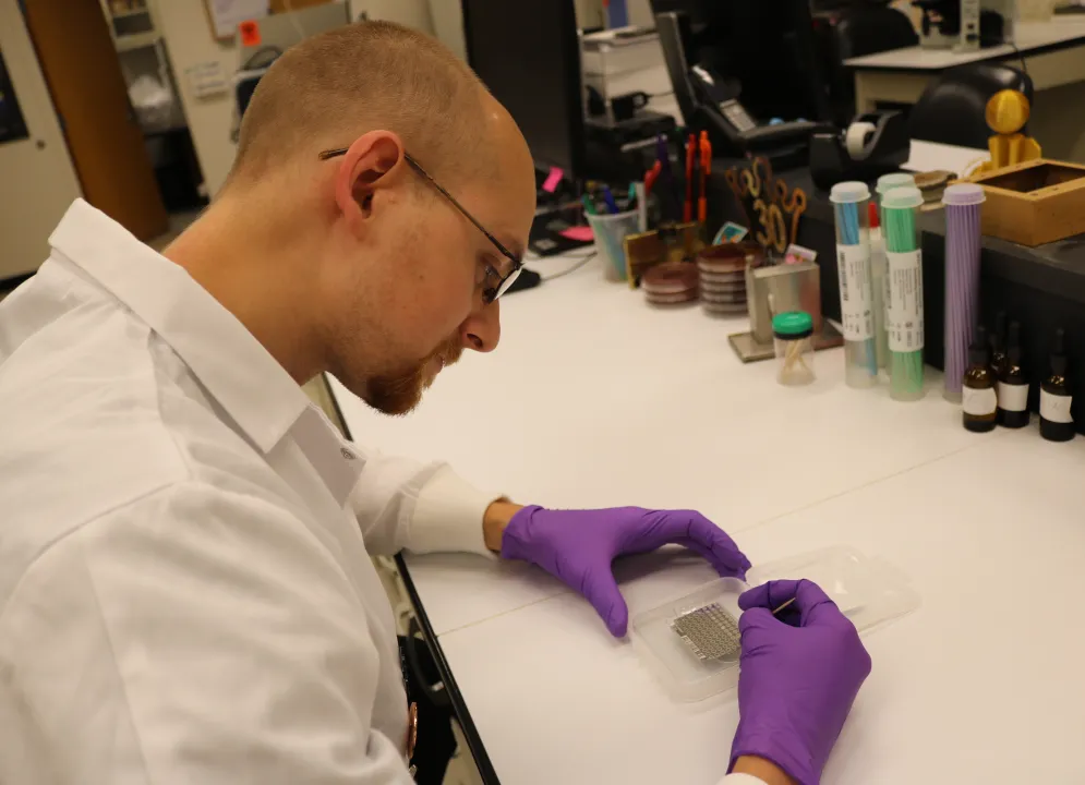
Comparative Pathology and Digital Imaging Shared Resource
Comparative Pathology and Digital Imaging Shared Resource (CPDISR) provides expert, readily available and affordable experimental pathology support to investigators utilizing animal models to study human disease.
What is Comparative Pathology and Digital Imaging Shared Resource?
Comparative pathologists affiliated with the CPDISR are familiar with normal anatomy and physiology, as well as background age- and strain-related lesions of various animal models.
Recognition of lesions and their interpretation in the context of individual investigations provides a critical component to research incorporating animal models.
Services include: comprehensive macroscopic and microscopic examinations of various species of laboratory animals with an emphasis on the phenotypic characterization of newly produced lines of genetically engineered mice.
Additional services include hematology, clinical chemistry, radiography; and, in conjunction with the Histology and Immunohistochemistry Core, routine frozen and paraffin slide preparation as well as tissue microarray preparation and special histochemical and immunohistochemical staining.
In addition, the CPDISR provides a referral service to experienced scientists within the Ohio State research community providing expertise in animal model development, experimental design, optimal sample collection and data interpretation.
Histology and Immunohistochemistry
Comparative Pathology and Digital Imaging Shared Resource also provides Histology and Immunohistochemistry services. The combined service offers anatomic pathology (necropsy, phenotyping, biopsy and slide evaluation), clinical pathology, and histology (including immunohistochemistry and tissue microarrays) support to researchers both within the College of Veterinary Medicine as well as throughout The Ohio State University, including Nationwide Children's Hospital.
Note: Services to non-University clients are by special prior arrangement.
Comparative and Clinical Pathology
Comparative and Clinical Pathology Services and Fees
- Fees: Current Fee Schedule (FY25)
- Info: CPDISR ERAMP Users Guide
Note: Fees are specifically for animal research pathology cases submitted to the Comparative Pathology & Digital Imaging Shared Resource.
CPDISR provides complete research animal services with knowledgeable staff and American College of Veterinary Pathologists board-certified comparative pathologists guiding the process. Our services include standard, customizable (comprehensive) and phenotyping necropsy services, biopsy services, slide evaluation services, and comprehensive clinical pathology for rodent specimens.
Our Necropsy and Biopsy bundle services include everything needed to go from live animal model or tissue sample to formal pathology evaluation by our board-certified comparative pathologists and their complete reports for an affordable price. To learn about what is included in these bundle services, please contact us at CPDISR@osu.edu
Comparative Pathology
Ideally, three to five mice of each genotype, including the same number of age- and sex-matched controls should be submitted for evaluation. Controls should be littermates exposed to the same environment and experimental conditions, and NOT mice of the same background strain purchased from a commercial vendor. After a brief ante mortem period of observation, the mice are euthanized by carbon dioxide asphyxiation, weighed and blood collected by percutaneous cardiac puncture for subsequent hematology (complete blood count including erythrocyte, leukocyte and platelet parameters with white blood cell differential) and clinical chemistry (29 routine serum assays evaluating liver and kidney function, electrolytes and protein levels). We have a network of reference laboratories available to provide additional tests that are not performed in-house. Urine and other fluids can also be analyzed; however, due to the small volumes typically obtained from mice, pooling of samples from mice of the same genotype, age, and sex may be indicated. Digital gross photographs are taken of any lesions in mutants and their lack thereof in controls.
A complete necropsy is performed. All organs are examined grossly, and the following organs are routinely weighed to the nearest milligram prior to fixation for determining organ-to-body weight ratios (% body weight):
Other organs can be weighed based on the pathologist's recommendation and/or the client's request.
Tissues are trimmed according to a standardized protocol, fixed in 10% neutral buffered formalin, processed by routine methods, embedded in paraffin wax and sectioned at 4 microns for standard H&E; unstained and frozen sections can also be made. All bony tissues are decalcified prior to trimming. The tissues noted in the table below are evaluated histologically by a comparative pathologist board-certified by the American College of Veterinary Pathologists (ACVP), frequently with the involvement of a veterinary pathology or laboratory animal trainee. Final reports include detailed findings, photomicrographs of relevant findings related to the phenotype, and an interpretative summary and recommendations for ancillary analyses offered by the CPDISR or other Shared Resources.
Block/Slide # |
|
| 1 |
|
| 2 |
|
| 3 |
|
| 4 |
|
| 5 |
|
| 6 |
|
| 7 |
|
| 8 |
|
| 9 |
|
| 10 |
|
| 11-12 |
|
| 13 |
|
| 14+ |
|
Comparative Pathology Frequently Asked Questions
In addition to phenotypic evaluations of genetically modified mice (GEM), the CPDISR offers standardized and customized necropsy and biopsy, trimming of tissue specimens for slide presentation, frozen (OCT) block preparation from fresh tissues, gross and microscopic digital photography and slide scanning, histopathological slide evaluation, pathology consultation, and training in related lab animal pathology techniques. Please see our services and fee schedule for details and contact us at CPDISR@osu.edu
Evaluations can be carried out in all veterinary species. Please contact our laboratory at 614-247-8122 or CPDISR@osu.edu PRIOR to the submission of live animals. Live animals must be submitted in covered cages. Call ahead for scheduling – we cannot accept unscheduled drop-offs! This is particularly important for live animals and blood/fluid samples.
For non-phenotpying cases involving live animals, fixed tissues or slides OR for submission of blood, serum or other fluids:
All service requests must be submitted through the OSU CCC eRAMP System (https://eramp.osumc.edu/welcome). Please contact our staff for assistance and utilize the CPDISR eRAMP Users Guide (link for download). After obtaining fiscal approval for your submission, arrange to drop off your samples or animals to the correct location in our Shared Resource:
Live Animals, Anatomic and Clinical Pathology Services: 471 Veterinary Medicine Academic Building (VMAB), 1900 Coffey Road.
Histology-only Services: 302 Goss Laboratory, 1925 Coffey Road
For phenotyping cases of previously uncharacterized genetically modified mice (GEMs):
Please download and complete our Phenotyping Pre-Submission Form (link to form) and email the competed form to CPDISR@osu.edu. A CPDISR pathologist will contact you to discuss your submission and assist with scheduling your phenotyping necropsies.
The requested information is essential for a valid pathological evaluation and interpretation. Published references pertaining to any genetic manipulation(s), treatment(s), or animal model(s) are particularly helpful. If you do not have the information on hand at submission, please provide this to your CPDISR comparative pathologist as soon as possible.
We can receive submissions from 8AM to 4PM Mon-Fri, unless alternative arrangements have been made. The CPDISR main lab is located in the Veterinary Medical Academic Building (VMAB), 1900 Coffey Road, room 471. The VMAB is a new 4-story building on the CVM campus in the cul-de-sac off Coffey Road with a flag pole at the main entrance. From the main entrance take the elevator to your right (west). Upon exiting the elevator on the 4th floor, you may obtain entrance to the lab by calling the lab at 614-247-8122. One of our technical staff will provide entry to the research corridor and assist you with your submission.
Please call the lab at 614-247-8122 as soon as possible to discuss. If there is insufficient time for proper processing that day, we may ask you to contact your ULAR veterinarian for euthanasia and/or necropsy assistance.
If an animal dies after hours or during a weekend, you should ideally perform a gross necropsy and preserve the tissues in formalin for submission during regular business hours; if needed, CPDISR technicians are available to train you in gross necropsy and tissue fixation techniques. Otherwise, immediately refrigerate – DO NOT FREEZE - the animal and submit the carcass on ice ASAP during regular business hours. Marked autolysis may preclude any definitive histological interpretation, so it is important to refrigerate or preserve dead animals as soon as possible.
Due to the variability of our workloads from week to week and the fact that we currently have only one dedicated veterinary pathologist, we cannot guarantee delivery times. In general, our anatomic cases are usually completed within 6-8 weeks. However, all reasonable efforts will be made to accommodate deadlines for publications, grant applications, or meetings. Please notify the CPDISR staff of such deadlines at the time of submission.
Unless otherwise requested, reports are delivered via the eRAMP Notes Section in pdf format. Accompanying digital photomicrographs and gross photographs will be provided via eRAMP or, if file sized are large, via OneDrive.
Yes. The CPDISR will not release any information about your specimen(s)/case(s) to any other individual without your express permission. Some selected specimens may be used in teaching veterinary pathology and laboratory animal residents, but those materials will not be used in scientific publications of any form without the permission of the owner of those specimens.
All glass slides, tissue blocks, wet tissues, etc., will be returned to the PI. You will be notified when your materials are available for collection at our main laboratory in 471 VMAB. If materials are not collected, CPDISR will hold the materials for 6 months, after which disposition as determined by the Director will be undertaken. Selected materials may be used for teaching veterinary pathology and laboratory animal residents. Wet tissue (that tissue remaining after trimming and slide generation) will be disposed of within a month of the case final report and billing unless the PI requests otherwise.
All OSU billing is done through OSUCCC eRAMP via WorkDay, via accounting services of The Ohio State University Comprehensive Cancer Center.
For non-OSU investigative faculty, a comparative pathologist will work with you to produce a formal quote that you may use to procure a Purchase Order (PO) prior to submitting your service request via eRAMP. We require upload of a PO to the Notes Section of a submission prior to initiation of work.
Clinical Pathology Services
Hematology is the study of the cellular components of blood, including red blood cells (RBCs), white blood cells (WBCs) and platelets The CPDISR offers:
- a complete automated blood count with WBC differential (CBC w/diff),
- a manual differential from a stained blood smear, and/or
- preparation of a blood smear, stained or unstained
A complete CBC w/differential requires a minimum of 30 μL of whole blood collected in an EDTA-coated tube (lavender top) at room temperature and submitted to the lab as soon as possible after collection to ensure valid results.
The complete blood count, or CBC, is performed on an automated analyzer and quantitates the cellular make-up of the blood in terms of the absolute and relative numbers (%) of RBCs (red blood cells), WBCs (white blood cells), and platelets per sample volume. Also included in the report is a more in-depth analysis of specific RBC and platelet indices such as PCV (packed cell volume or hematocrit), hemoglobin, and MCV (mean corpuscular volume), among others. Abnormal values are frequently indicative of disease processes, although values can also differ according to species, strain, sex and age as well as to husbandry conditions and stress.
Manual differentials are performed by a trained medical technician on stained blood smears to complement the automated CBC. Manual evaluation of blood smears are performed to ensure correlation with automated counts and to detect morphological cellular changes such as abnormal shapes, structural remnants, parasites or viral inclusions.
Blood smears are prepaired manually, air-dried, and stained with standard Wright Giemsa.
Serum chemistry, or clinical chemistry, is the analysis of the blood serum or plasma for levels of electrolytes, enzymes, lipids, carbohydrates, proteins, blood gases, and metabolic products. The process is automated. As with the CBC, abnormal values are frequently indicative of disease processes, although values can also differ according to husbandry conditions, stress, species, strain, sex, and age.
The CPDISR offers several standard panels which include:
Comprehensive profile
- albumin
- alkaline phosphatase (ALP)
- alanine aminotransferase (ALT)
- amylase
- aspartate aminotransferase (AST)
- blood urea nitrogen (BUN)
- calcium
- chloride
- cholesterol
- creatinine
- creatinine kinase (CK)
- gammaglutamyl transferase (GGT)
- globulins
- glucose
- lipase
- phosphorus
- potassium
- sodium
- total bilirubin
- total protein
- trigylceride
Cardiac/lipid panel
- cholesterol (total)
- lactate dehydrogenase (LDH)
- HDL and/or LDL
- creatinine kinase (CK)
- triglyceride
Electrolytes
- chloride
- potassium
- sodium
Renal panel
- albumin
- globulin
- glucose
- blood urea nitrogen (BUN)
- phosphorus
- calcium
- potassium
- chloride
- sodium
- cholesterol
- total protein
- uric acid
- creatinine
Hepatic panel
- albumin
- alkaline phosphataes (ALP)
- alanine aminotransferase (ALT)
- aspartate aminotranferase (AST)
- direct bilirubin
- indirect bilirubin
- total bilirubin
- gamma glutamyl transferase (GGT)
- globulin
- total protein
Single tests can also be ordered and any of the above panels can be adjusted to the individual needs of the PI. We encourage prompt submission of sera, but serum refrigerated for a short period of time or frozen may still yield valid results for some tests.
Urinalysis is the evaluation of urine collected by free-catch, manual expression, or cytocentesis by dip stick for several parameters such as pH and metabolic products, by refractometry for specific gravity, and by visual inspection of sediment. Ideally, urine is collected in sterile receptacles and submitted as soon as possible, but, if necessary, clean urine can be stored refrigerated overnight. Degradation increases over time and may adversely influence the analysis results, even with refrigeration; freezing is discouraged if cellular elements are an important factor. Although approximately 1 ml of clean urine is required for complete analysis, selected parameters can be tested on smaller volumes. If necessary, urine may be pooled from multiple mice providing they are of the same strain and genotype, age, sex, and treatment (if applicable).
Bronchioalveolar lavage (BAL)/fluid analysis is an examination of the cellular components of fluids collected from tracheal washes, pleural effusions, abdominal ascites, cystic structures, etc. Automated cell counts and/or cytospins are stained to delineate and characterize cellularity. Fluid analysis requires about 1 ml of sample for accurate interpretation, and, while fresh specimens are preferred, samples refrigerated overnight may still yield valid results.
Cytology is the assessment of cells collected by aspiration and swabs, as well as slide smears or imprints, from superficial or internal tissues or lesions and typically complements a fluid analysis. Cytological specimens are stained and evaluated by light microscopy.
Clinical Pathology Frequently Asked Questions
The CPDISR currently offers hematology, serum biochemistry, analysis of urine and other body fluids, and cytology. All tests are performed by qualified laboratory professionals trained in veterinary laboratory techniques. The laboratory staff is committed to research in improved diagnostics, reference range development, and state-of-the-art assays; new tests are continually being added as experimental needs change. We also have a network of reference laboratories available to provide tests that are not currently performed in-house.
We can assay samples from a wide variety of species, including some exotics, and from virtually all large and small laboratory animals.
Please see our lists of tests in the Services [link to main clinical path page] section. If you have questions or do not see a desired assay, please contact the lab at 614-247-8122 or CPDISR@osu.edu. Please see our current fee schedule [Link to pdf] for price per test.
Sample preparation is dependent on the tests you wish to run – please contact us for assistance! To help us provide accurate and reliable results, please contact us at 614-247-8122 or CPDISR@osu.edu PRIOR to sample collection and submission.
Submission of service requests is completed through OSU CCC eRAMP System (https://eramp.osumc.edu/welcome). Please contact our staff for assistance and utilize the CPDISR eRAMP Users Guide (link for download). After obtaining fiscal approval for your submission, arrange to drop off your samples or animals to the correct location in our Shared Resource:
471 Veterinary Medicine Academic Building (VMAB), 1900 Coffey Road.
The clinical pathology lab is open from 8 a.m. to 5 p.m. Monday-Friday; however, samples should be submitted before 1 p.m., unless previous arrangements have been made, just to ensure that your tests can be run that same day. Whole blood must be submitted as soon after collection as possible. If you anticipate submitting a large number of samples (> 25), please call the lab one or two days prior to submission to facilitate scheduling.
Hematology, serum chemistry and urinalysis results are usually forwarded to you within 48 hours via the eRAMP Notes Section. Analysis of blood smears and cytology submissions may require up to 5 working days as a manual observation is performed by our med tech to confirm the automated counts and to detect changes in cellular morphology. If submitted as stand-alone samples, the results will be scanned and emailed to you, unless you make arrangements to pick them up from the CPDISR lab. The results of clinical pathology performed in conjunction with anatomic pathology submissions are correlated with the histopathological findings and provided by the pathologist at case completion.
All OSU billing is done through OSUCCC eRAMP via WorkDay, via accounting services of The Ohio State University Comprehensive Cancer Center.
For non-OSU investigative faculty, a comparative pathologist will work with you to produce a formal quote that you may use to procure a Purchase Order (PO) prior to submitting your service request via eRAMP. We require upload of a PO to the Notes Section of a submission prior to initiation of work.
Slide Scanning and Image Analysis
- Fees: Current Fee Schedule (FY23)
- Info: CPDISR ERAMP Users Guide
CPDISR offers Whole Slide Scanning and Image Analysis capabilities.
For Slide Scanning via the Animal Arm of CPDISR, an Olympus Whole Slide Scanner capable of Brightfield and Fluorescent scanning is housed in Goss Laboratory. Investigators may request slide scanning as part of their eRAMP submissions for histology or anatomic pathology services. For assistance with submission of slide scanning requests, see the CPDISR eRAMP Users Guide and contact our staff at 614-247-8122 and CPDISR@osu.edu.
For Slide Scanning and Image Analysis through the Digital Imaging portion of our Shared Resource, including the services of an Image Analysis specialist, contact our staff at CPDISR@osu.edu.
Contact
CPDISR is available by contacting:
Kara Corps, DVM, PhD
Director
Phone: 614-292-4261
Email: corps.2@osu.edu
Comparative Pathology & Digital Imaging Shared Resource
The Ohio State University College of Veterinary Medicine
Department of Veterinary Biosciences
Veterinary Medicine Academic Building 467/471
1900 Coffey Road
Columbus, OH 43210
Phone: 614- 247-8122
Email: CPDISR@osu.edu